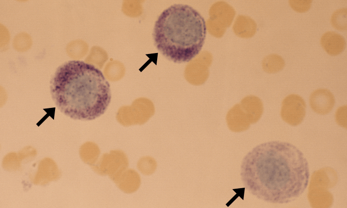
Mast Cells

Mast Cells 3 (Canine 3 - Mast Cell Leukemia) ARROWS
Mast cells. Canine (mast cell leukemia).

Morphology: leukocyte with a large round nucleus and numerous dark purple or blue cytoplasmic granules. Usually seen at the feathered edge of blood smear. Considerable variation in appearance of mast cells.

Look alikes: other leukocytes with cytoplasmic granules (basophils, eosinophils)

Clinical relevance: not present in the blood of healthy animals. In dogs, mastocytemia (↑ mast cells in peripheral blood) may be associated with neoplasia or with non-neoplastic conditions such as allergic reactions and severe inflammation (IMHA, pancreatitis, peritonitis, pleuritis, parvoviral enteritis). In cats, the presence of mast cells in the blood is almost always associated with neoplasia. Neoplastic mastocytemia may occur secondary to solid mast cell tumors (visceral or cutaneous) or due to primary bone-marrow-derived mast cell leukemia.

Mast Cells 1 (Canine 1 - Mast Cell Leukemia) ARROWS
Mast cells (arrows). Neutrophil (arrowhead). Canine (mast cell leukemia).
Mast Cells 2 (Canine 2 - Mast Cell Leukemia) ARROWS
Mast cells (arrows). Lymphocyte (arrowhead). Canine (mast cell leukemia).
Mast Cells 6 (Canine 4 - Mast Cell Leukemia) ARROWS
Mast cell (arrow). Two neutrophils and one lymphocyte (arrowheads) also visible. Canine (mast cell leukemia).
Mast Cells 7 (Canine 5 - Mast Cell Leukemia) ARROWS
Mastocytemia. Numerous mast cells (arrows). Numerous neutrophils and one lymphocyte also visible (not marked). Canine (mast cell leukemia).
Mast Cells 4 (Feline 1) ARROWS
Mast cells. Feline.
Mast Cells 5 (Feline 2 - Mast Cell Leukemia) ARROWS
Mast cell. Feline (mast cell leukemia).


Leave a Reply

Fill in your details below or click an icon to log in:

WordPress.com Logo

You are commenting using your WordPress.com account. Log Out /  Change )

Facebook photo

You are commenting using your Facebook account. Log Out /  Change )

Connecting to %s