Spherocytes

Spherocytes 3 (Canine 2 - IMHA) ARROWS
Numerous spherocytes (examples marked with arrows). Normal red blood cell (arrowhead) for comparison. Canine (IMHA).

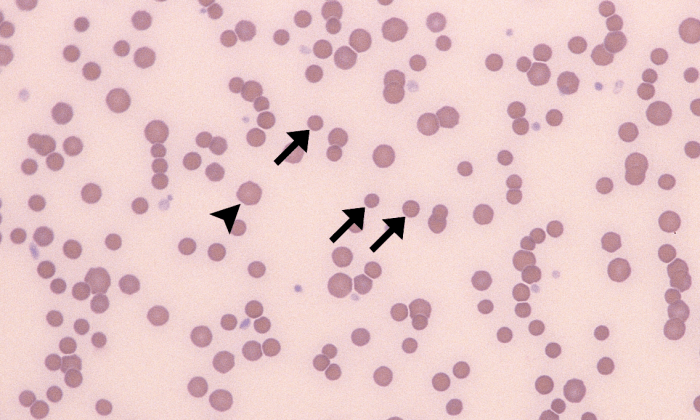
Morphology: red blood cells with a spherical rather than biconcave shape. Spherocytes appear smaller than normal red blood cells, with loss of central pallor in dogs.

Look alike: microcytes (may be difficult to distinguish microcytes from spherocytes in cats, horses, and cattle due to lack of central pallor in normal red blood cells)

Commonly seen with: other features of immune-mediated hemolytic anemia (IMHA) including ghost cells, polychromatophils, and agglutination of red blood cells

Clinical relevance: large numbers of spherocytes are associated with IMHA. Spherocytes may also be observed in stored or transfused blood. Very small numbers of spherocytes in the feathered edge may not have any clinical significance. Presence of spherocytes does not usually affect mean cell volume (MCV) on CBC because cell volume is the same (cell has decreased diameter but increased roundness).

Spherocytes 2 (Canine 1 - IMHA) ARROWS
Numerous spherocytes (examples marked with arrows). Normal red blood cell (arrowhead) for comparison. Lymphocyte and neutrophil also visible (not marked). Canine (IMHA).
Spherocytes 1 (Equine 1 - IMHA) ARROWS
Numerous suspected spherocytes (examples marked with arrows). Normal red blood cell (arrowhead) for comparison. Equine (IMHA).
Spherocytes 4 (Feline 1 - IMHA) ARROWS
Numerous suspected spherocytes (examples marked with arrows). Normal red blood cell (arrowhead) for comparison. Polychromatophils, ghost cells, and platelets also visible (not marked). Feline (IMHA).

Leave a Reply

Fill in your details below or click an icon to log in:

WordPress.com Logo

You are commenting using your WordPress.com account. Log Out /  Change )

Facebook photo

You are commenting using your Facebook account. Log Out /  Change )

Connecting to %s