Reticulocytes
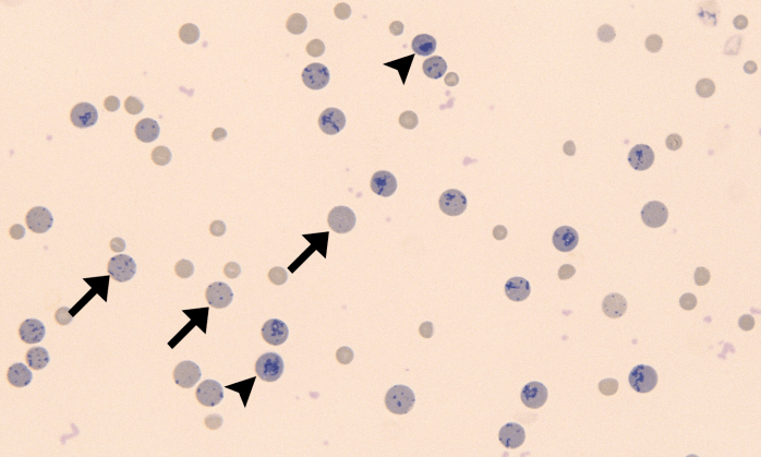
Morphology: immature red blood cells that stain blue with new-methylene blue due to precipitation of retained cytoplasmic RNA. Aggregate reticulocytes are less mature with large amounts of RNA that form dark blue clumps or strands. Punctate reticulocytes are more mature with small amounts of RNA that form small blue dots.
Commonly seen with: features of regenerative anemia (macrocytosis, anisocytosis, polychromatophils, nucleated red blood cells)
Polychromatophils versus reticulocytes: both are immature red blood cells containing RNA. Reticulocytes stain blue with new-methylene blue. Polychromatophils stain purple with Romanowsky stains. Aggregate reticulocytes stain purple on Romanowsky stains stains so aggregate reticulocytes are also polychromatophils. Punctate reticulocytes cannot be distinguished from normal red blood cells on Romanowsky stains.
Clinical relevance: increased reticulocytes (reticulocytosis) indicates that bone marrow is responding to anemia. If a robust polychromatophilic response is not present in an anemic patient, it is important to evaluate a blood smear using new-methylene blue stain to detect reticulocytes. Anemia is considered regenerative if sufficient reticulocytes are present. A reticulocyte count (percentage or absolute count) quantifies the number of aggregate reticulocytes present (punctate reticulocytes are not included in the reticulocyte count because they are more mature and do not reflect current regeneration).


