Monocytes

Monocytes 1-3 (Canine 1-3) ARROWS
Monocytes showing variable morphology and pleomorphic nuclei. Canine.

Morphology: leukocytes with pleomorphic nuclei (usually lobulated but may be round or kidney-shaped) and blue-gray cytoplasm containing occasional vacuoles or fine pink granules. Monocytes are larger than most other leukocytes. Considerable variation in appearance within and between species.

Look alike: band neutrophils (canine monocytes may have band-shaped nuclei), lymphocytes (equine monocytes may resemble lymphocytes).

Clinical relevance: monocytosis (↑ monocytes in peripheral blood) may be associated with corticosteroids (in dogs), granulomatous inflammation, immune-mediated diseases, tissue necrosis, and neoplasia. Monocytes leave the blood and transform into macrophages within tissues, so significant inflammation or immune-mediated disease may be present without causing monocytosis.

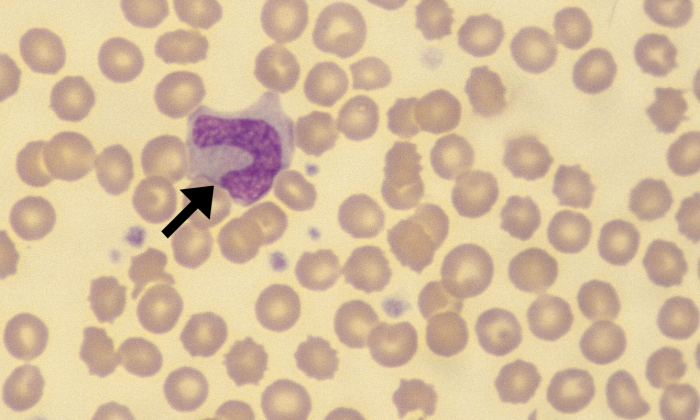
Monocytes 6-7 (Canine 6-7) ARROWS
Monocytes showing variable morphology, cytoplasmic vacuoles, and pleomorphic nuclei. Canine.
Monocytes 4 (Canine 4) ARROWS
Monocyte with band-shaped nucleus. Canine.
Monocytes 8 (Canine 8) ARROWS
Monocytes with kidney-shaped nucleus (top) and band-shaped nucleus (bottom). Canine.
Monocytes 5 (Canine 5) ARROWS
Monocyte. Canine.
Monocytes 9 (Canine 9) ARROWS
Monocytes. Cytoplasmic vacuoles visible in monocyte on right. Canine.
Monocytes 10 (Bovine1) ARROWS
Monocyte with cytoplasmic vacuoles. Bovine.
Monocytes 11 (Goat 1) ARROWS
Monocytes (arrows). Neutrophils (arrowheads). Lymphocyte (circle). Dall’s Sheep.

Leave a Reply

Fill in your details below or click an icon to log in:

WordPress.com Logo

You are commenting using your WordPress.com account. Log Out /  Change )

Facebook photo

You are commenting using your Facebook account. Log Out /  Change )

Connecting to %s